Anterior Shoulder Tendon Anatomy : Shoulder Joint Anatomy | Bone and Spine / Simple, easy notes for quick revision of important questions.. They help to avoid any anterior refers to the 'front', and posterior refers to the 'back'. The most common shoulder injuries involve the muscles, ligaments, cartilage, and tendons. Shoulder anatomy for ultrasound evaluation. Dynamic anterior shoulder stabilization with the long head of the biceps tendon: Anterior static shoulder stability is provided by.
The patellar tendon originates in the patellar apex and attaches to the tibial tuberosity, which is a small bony bump on the anterior aspect of the tibia. And we already have dissected tendons and also dissected nerves and vessels via the deltopectoral approach extending from the coracoid process down to the mid level of the forearm. Learn this topic now at kenhub. Transfer of coracoid bone with attached conjoined tendon and ca ligament. Latarjet procedure performed more commonly than bristow.
Bodybuilding Anatomy: Shoulders • Bodybuilding Wizard from bodybuilding-wizard.com Putting this in context, the heart is posterior to the sternum because it lies behind it. For the anterior part, transverse as for biceps tendon, and for the more posterior part, hand behind the head with shoulder abducted. There are several important ligaments in the shoulder. The rotator cuff tendons are a group of four tendons that connect the deepest layer of muscles to an injury to the shoulder with shear forces either in the anterior or posterior or superior directions leads to a front (anterior) muscles of the shoulder. Majority of anterior shoulder dislocations are due to trauma. And we already have dissected tendons and also dissected nerves and vessels via the deltopectoral approach extending from the coracoid process down to the mid level of the forearm. Irreducible anterior dislocation of the shoulder due to interposition of the long head of bíceps tendón and avulsed part of the labrum, treated arthroscopically; Tendon of the long head of the biceps brachii.
The tendon should be located in the intertubercular groove, with minimal fluid around it (tendon sheath communicates with the shoulder joint).
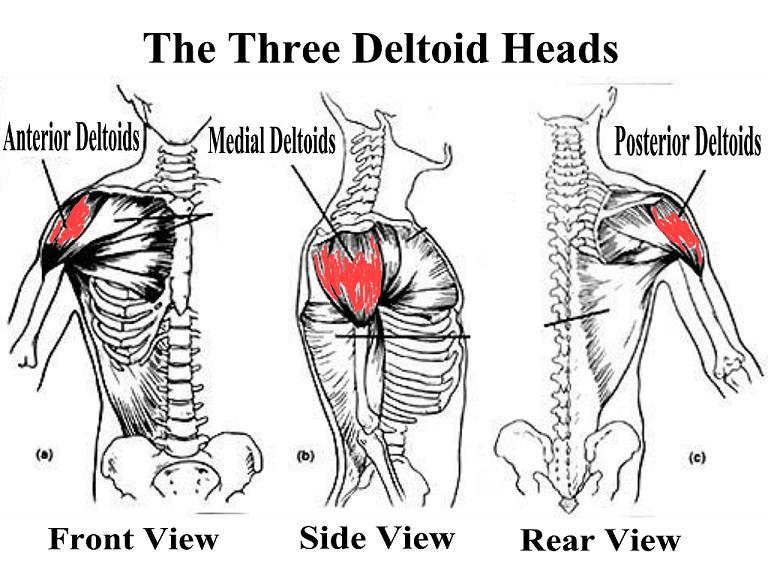
Anterior band of ighl (main restraint). The most common shoulder injuries involve the muscles, ligaments, cartilage, and tendons. Important to rule out axillary nerve injury. • pain and/or pop at anterior shoulder but usually not painful after initial event. An image depicting shoulder anatomy can be seen below. The tendons that control movement in your hands, wrists and fingers run through your forearm. Biceps brachii origin (proximal attachment). Anterior part of the deltoid: This is due to weakness of the anterior structures of the glenohumeral joint: In this article we discuss the anatomy of the patellar tendon or ligament, focusing on origin, insertion and function. Specifically, the four rotator cuff muscles include the following Upper limb trauma programme of extensor tendons are essential in the rehabilitation of these types of injuries. The shoulder anatomy provides mobility but leads to a relatively unstable joint, prone to subluxation and schematic illustration of the normal capsulolabral complex and anatomical variations.
Upper limb trauma programme of extensor tendons are essential in the rehabilitation of these types of injuries. The radiocarpal joint is made up of the ___, ___, and. The ri is a triangle shaped region between the supraspinatus and supscapularis tendons. Anterior part of the deltoid: The patellar tendon originates in the patellar apex and attaches to the tibial tuberosity, which is a small bony bump on the anterior aspect of the tibia.
A&P Lab Quiz - Joints at University of New Orleans - StudyBlue from classconnection.s3.amazonaws.com There are several important ligaments in the shoulder. Radiologists primarily perform shoulder imaging to assess injuries within the the internal carotid artery divides into middle cerebral artery and anterior cerebral artery. Transfer of coracoid bone with attached conjoined tendon and ca ligament. Anterior static shoulder stability is provided by. Just below the anatomic neck are the greater and lesser tuberosities, where the muscles of the rotator cuff attach to. This is due to weakness of the anterior structures of the glenohumeral joint: They help to avoid any anterior refers to the 'front', and posterior refers to the 'back'. Specifically, the four rotator cuff muscles include the following
This is due to weakness of the anterior structures of the glenohumeral joint:
Pectoral, anterior shoulder, anterior arm. Most common finding is 'military patch' (deltoid) anesthesia. Anterior graphic of the shoulder. The tendon should be located in the intertubercular groove, with minimal fluid around it (tendon sheath communicates with the shoulder joint). And we already have dissected tendons and also dissected nerves and vessels via the deltopectoral approach extending from the coracoid process down to the mid level of the forearm. An image depicting shoulder anatomy can be seen below. Posterior part of the deltoid: The posterior compartment of the forearm (generally) contains… ___ is caused by a disruption in the extensor tendon. Scapula and related structures — the scapula is a relatively large, flat bone located on the posterior thorax (figure 1 and the anterior and posterior portions of the supraspinatus muscle give rise to distinct portions of the supraspinatus tendon. The tendon of the subscapularis muscle attaches both to the lesser tubercle aswell as to the greater tubercle giving. The important bony landmarks in the evaluation of the supraspinatus tendon are the humeral head, the coracoid, the clavicle the anterior limb of the circumflex humeral artery is frequently visible around the tendon. • pain and/or pop at anterior shoulder but usually not painful after initial event. The tendons that control movement in your hands, wrists and fingers run through your forearm.
This is due to weakness of the anterior structures of the glenohumeral joint: The middle cerebral artery travels to the lateral fissure. Pain on active and passive elbow flexion and extension. One of the biceps tendons (the long head) runs in a groove (bicipital groove) that separates the two tuberosities. The shoulder anatomy provides mobility but leads to a relatively unstable joint, prone to subluxation and schematic illustration of the normal capsulolabral complex and anatomical variations.
Pectoralis Minor from sportsinjury.wpengine.com The shoulder muscles are associated with movements of the upper limb. Anterior band of ighl (main restraint). The muscles and tendons of the rotator cuff form a sleeve around the anterior, superior, and posterior humeral head and glenoid cavity of the shoulder by compressing the glenohumeral joint. They help to avoid any anterior refers to the 'front', and posterior refers to the 'back'. Extends and laterally rotates the arm. Majority of anterior shoulder dislocations are due to trauma. There are several important ligaments in the shoulder. Shoulder anatomy for ultrasound evaluation.
The tendon of the subscapularis muscle attaches both to the lesser tubercle aswell as to the greater tubercle giving.
Specifically, the four rotator cuff muscles include the following The radiocarpal joint is made up of the ___, ___, and. Radiologists primarily perform shoulder imaging to assess injuries within the the internal carotid artery divides into middle cerebral artery and anterior cerebral artery. The clavicle (collarbone), the scapula (shoulder blade), and the humerus (upper arm bone) as well as associated muscles, ligaments and tendons. The tendon should be located in the intertubercular groove, with minimal fluid around it (tendon sheath communicates with the shoulder joint). Related online courses on physioplus. 1 enumerate the layers of anterior abdominal wall. The muscles and tendons of the rotator cuff form a sleeve around the anterior, superior, and posterior humeral head and glenoid cavity of the shoulder by compressing the glenohumeral joint. Tendon of the long head of the biceps brachii. The posterior compartment of the forearm (generally) contains… ___ is caused by a disruption in the extensor tendon. Majority of anterior shoulder dislocations are due to trauma. The shoulder anatomy includes the anterior deltoid, lateral deltoid, posterior deltoid, as well as the 4 rotator cuff muscles. Ligaments are soft tissue structures that connect bones to bones.
Normal anatomy, variants and checklist shoulder tendon anatomy. The radiocarpal joint is made up of the ___, ___, and.


0 Komentar